

A child sits in the dental chair, a parent watches the sensor being prepared, and one question comes quickly: “Is this really safe?” That hesitation is understandable. Dental X-rays use ionizing radiation, so families in Amanda, Lancaster, Circleville, and Carroll, Ohio, deserve a clear explanation before any image is taken.
Dental X-ray safety isn't about avoiding every image. It's about taking the right image only when it can answer an important clinical question, using the lowest practical exposure, and avoiding unnecessary repeats. For patients looking for a dentist in Amanda, OH, a family dentist near me, or an emergency dentist after tooth pain, that distinction can make diagnostic care feel far less intimidating.
Table of Contents
- Understanding Dental X-Ray Safety for Your Family
- Comparing Dental Radiation to Everyday Exposure
- How Digital Technology Reduces Your Exposure
- Special Considerations for Pregnancy and Children
- Why Skipping Necessary Images Carries Greater Risk
- Your Comfort and Care at Amanda Family Dental
Understanding Dental X-Ray Safety for Your Family
A child opens wide, the sensor comes out, and a parent wonders whether the image is really needed. That moment matters. At Amanda Family Dental, the first step is not taking an X-ray by default. It is deciding whether the image will answer a question that a regular exam cannot.
Some dental problems stay hidden from view. A dentist may need an image to check for decay between teeth, infection near a root, bone changes, or a tooth that is still trapped under the gums. Those findings can change what happens next, whether that means a filling, root canal, tooth extraction, restorative dentistry, or careful monitoring.
A useful way to judge dental X-ray safety is to focus on the decision, not just the machine. The standard for responsible imaging is the ALARA principle, or “as low as reasonably achievable.” In plain language, that means using an image only when it can affect care, choosing the fewest images that still answer the clinical question, selecting settings that fit the patient and the job, and keeping equipment in proper working order. FDA's guidance on dental radiographic examinations identifies 60 to 70 kVp as the optimal operating range for dental X-ray units and advises against settings above 90 kVp.
A practical question before imaging
Patients and parents can ask for the reason behind a recommended image. A short conversation often makes the decision much less stressful:
- Clinical question: What problem is the image checking?
- Recent records: Are images from another office available?
- Treatment impact: Would the result change the recommended care?
- Exposure choice: Is a standard image enough, or is a larger scan being considered?
For example, a parent can ask, “We had bitewings six months ago at our previous office. Do you need new ones today, or can you request those records first?”
Patients can also review how often dental X-rays may be needed to see why imaging frequency depends on personal risk, symptoms, and findings rather than one routine schedule for every family.
Comparing Dental Radiation to Everyday Exposure
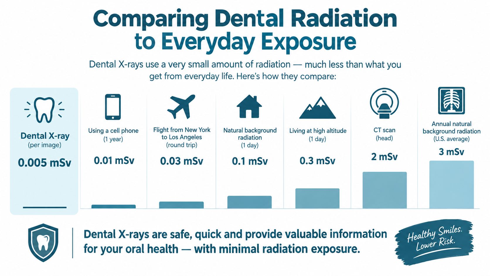
A parent often hears “radiation” and pictures something large and dangerous. In dentistry, the better question is simpler: what image is being recommended, and does it fit the problem we are trying to solve?
Dose terms such as mSv and µSv can feel abstract. They become easier to understand when you compare one dental image with other familiar exposures and, when you remember that different dental images are not all the same. The goal is not to chase the smallest possible number in every situation. The goal is to choose the smallest image that still answers the clinical question.
Published comparisons from the FDA show that a conventional dental image represents a relatively low level of exposure in relation to other common medical imaging examples, and the agency's public materials also make clear that “low” does not mean zero. That is why the decision still matters, even when the dose is small. An infographic comparing the low radiation dose of a dental x-ray to various common daily life activities.
A 2020 assessment by the United Nations Scientific Committee on the Effects of Atomic Radiation reported a typical effective dose of about 0.006 mSv for a dental intraoral examination, the lowest typical dose among the examination categories it evaluated. That does not make every visit identical, and it does not justify taking images that are not needed. It does give families a calmer, more realistic sense of scale.
The imaging type matters
A routine intraoral image and a cone-beam computed tomography scan, or CBCT, serve different purposes. Published IAEA training data report typical effective doses of approximately 0.3 to 21.6 µSv for an intraoral radiograph, 2.7 to 38 µSv for panoramic imaging, and 11 to 1,025 µSv for CBCT. The FDA also explains that CBCT usually exposes patients to more radiation than conventional dental X-rays, while remaining below many medical CT examinations. The FDA's CBCT guidance explains why the scan should answer a question that lower-dose imaging cannot answer.
For a patient in Lancaster, Circleville, or Carroll, the practical checklist is straightforward: What is the dentist looking for? Will a small image answer it? Is a panoramic view needed? Or does the situation really call for three-dimensional detail? The modality should match the clinical question.
Practical rule: A larger or more detailed scan is not automatically a better scan. The best image is the one that gives enough information for the decision being made.
How Digital Technology Reduces Your Exposure
A parent often hears, “We'll take a digital X-ray,” and the next question is simple: does digital really mean less exposure? In practice, it can, but the answer is not just the sensor. It is the whole process, choosing the right image, using the right settings, and avoiding retakes.
Older film and modern digital receptors are not handled the same way. In an FDA-reported 1999 survey, typical exposure per film was 1.7 mGy with D-speed film and 1.3 mGy with E-speed film. Switching from D-speed to E-speed film represented a 23% reduction, while moving from E-speed to F-speed film reduced exposure by another 20 to 25%. Changing directly from D-speed to F-speed film produced an approximately 60% reduction without compromising diagnostic quality. These figures come from the FDA's selection guidance for dental radiographic examinations. While those film-speed figures are historical, they help explain why current digital sensors are paired with collimation and careful exposure selection to keep dose as low as reasonably achievable.
Digital receptors support low-dose imaging in another way. As explained in this overview of digital X-ray advantages, the image is captured electronically, so the team can review it right away and confirm whether it answers the clinical question. That matters because a clear first image lowers the chance that a child or adult needs a repeat because of positioning or an exposure setting that missed the mark.
Four controls work together
Digital equipment works a bit like using a phone camera in low light. A better device helps, but the result still depends on aim, settings, and whether you are taking the right picture in the first place. Safe dental imaging follows that same checklist.
- Beam collimation: The beam is limited to the size of the receptor instead of exposing more tissue than needed.
- Exposure selection: Time and intensity are chosen for the patient and the job the image needs to do.
- Positioning: The sensor and patient are aligned carefully to reduce distortion and prevent retakes.
- Equipment care: Regular maintenance helps the unit produce dependable images at suitable settings.
Children need extra attention. They are more sensitive to ionizing radiation, and they have more years ahead in which potential effects could appear. That is why child-sized settings, careful positioning, and taking only the images needed for diagnosis matter so much.
Amanda Family Dental offers digital dental X-rays as part of its broader dental care approach. Patients can ask how a proposed image supports cleaning and exams, restorative dentistry, dental implants, or an urgent evaluation for tooth pain.
Why CBCT is different
CBCT can be useful when three-dimensional anatomy changes treatment planning, including selected implant or surgical cases. A conventional image should still be used when it can answer the question. The smallest appropriate field of view and image quality that is adequate for diagnosis keep exposure matched to the clinical need.
Special Considerations for Pregnancy and Children
Pregnancy often makes a routine dental decision feel urgent. A patient may worry that any X-ray could harm an unborn child, while postponing care could allow an infection or severe tooth pain to worsen. The Health Physics Society states that a complete dental X-ray examination produces an insignificant fetal dose and identifies no evidence of harm to an unborn child. Its guidance notes that conventional dental imaging has not been shown to pose a fetal risk. The Health Physics Society's dental patient guidance provides that context.
Patients should still tell the dental team about a pregnancy or possible pregnancy. The clinician can review previous images, decide whether the image changes immediate care, and select the lowest exposure compatible with diagnosis. More recent professional guidance reports fetal doses below 10 microgray for dental X-rays of the teeth and jaws, including CBCT, and does not recommend lead aprons specifically for dental imaging. Policies may differ by jurisdiction or practice, so patients should ask how the office handles protection.
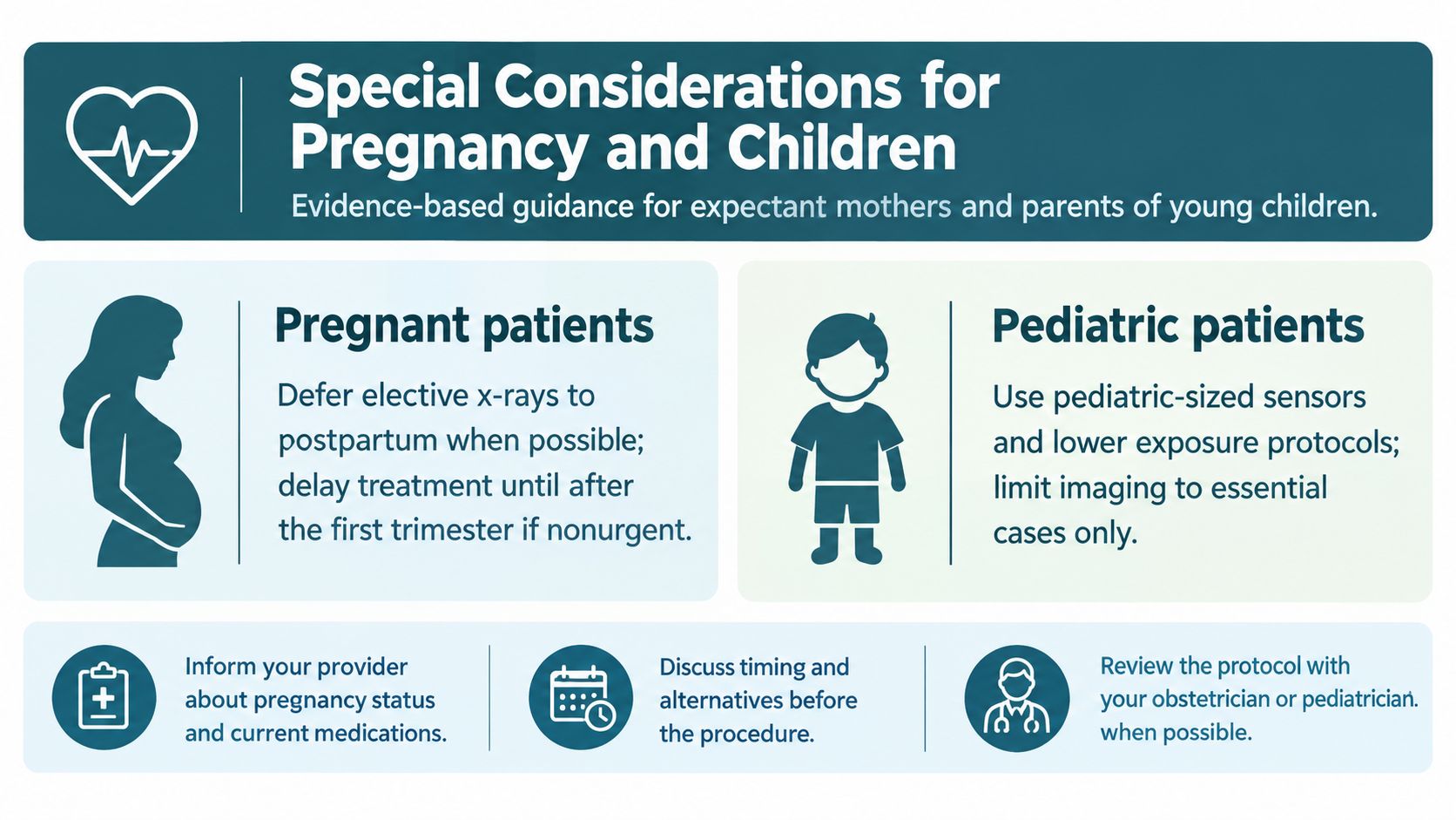
A pregnancy conversation can follow four steps
- Share the pregnancy status. The dental team needs accurate information before selecting imaging.
- Review existing records. Recent images may prevent an unnecessary repeat.
- Ask about urgency. A painful infection, swelling, or emergency may require timely diagnosis, while elective imaging may be considered differently.
- Discuss coordination. Patients can review the plan with an obstetrician when appropriate, especially if a broader scan is being considered.
Expectant parents may also benefit from practical essential prenatal preparation tips, including preparation for healthcare conversations and appointments.
Children need child-sized care
A child's smaller body and developing tissues make exposure settings especially important. The team should use the smallest practical sensor, child-appropriate exposure settings, precise beam restriction, and only the images needed to answer the diagnostic question. Parents can learn more about what to expect from a pediatric dental exam.
A child who has facial swelling, persistent tooth pain, an injury, or a developing tooth concern may need imaging because the visible symptom doesn't show the full problem. The goal isn't to expose a child “just in case.” The goal is to avoid guessing when an image can guide safer, more focused care.
Why Skipping Necessary Images Carries Greater Risk
Radiation anxiety can draw attention away from another risk: missing disease that can't be seen during a visual examination. An X-ray may show decay between teeth, infection near a root, bone changes below the gumline, or a tooth developing in an unusual position. Without that information, a dentist may not know whether a sore tooth needs a filling, root canal, extraction, or observation.
That matters because untreated disease can advance without notice. A small area of decay may become a larger restoration. An infection may develop into swelling or an urgent dental problem. An impacted tooth may complicate planning for orthodontic care, surgery, or a future dental extraction.
The decision is about benefit and necessity
A responsible clinician doesn't order imaging because a calendar says it's time. The decision considers symptoms, examination findings, age, oral health history, previous images, and whether the result will change care. The EPA describes a typical dental X-ray effective dose of about 0.04 mSv, while the Health Physics Society says a full-mouth series is generally below one month of natural background exposure. The EPA's radiation medicine information places dental imaging within a broader discussion of exposure and risk.
A 2025 systematic review cautions that repeated low-dose exposures accumulate over a lifetime and reinforces ALARA, especially for children and pregnant patients. That doesn't create a universal annual quota. It supports practical habits that prevent needless repeats:
- Bring prior images: Ask a previous office to send diagnostically adequate records.
- Ask what the image answers: A clear clinical reason should exist before exposure.
- Separate imaging types: Conventional images and CBCT carry different doses and purposes.
- Review alternatives: If an existing image is adequate, repeating it may not add value.
For families seeking a dentist near me in Amanda, Lancaster, Circleville, or Carroll, necessary imaging can protect comfort, function, and treatment options. Finding hidden disease early may help patients avoid more extensive restorative dentistry, emergency care, or tooth extraction later.
Your Comfort and Care at Amanda Family Dental
A new patient exam should feel like a conversation, not a conveyor belt. Patients can expect a review of their concerns, health history, and previous dental records before the team decides whether digital X-rays are needed. When imaging is appropriate, the sensor is positioned gently, exposure settings are selected for the patient, and the reason for the image is explained in plain language.
The practice provides preventive cleaning and exams, pediatric care, fillings, crowns, root canals, cosmetic dentistry, teeth whitening, Invisalign, dentures, dental implants, and emergency dental services. Patients searching for dental implants near me, a cosmetic dentist near me, or help with tooth pain can discuss how imaging supports a personalized treatment plan.
Amanda Family Dental also offers mercury-free options, fluoride-free choices, digital X-rays, total health screening, myofunctional therapy, and sleep dentistry. For patients who feel nervous, transparent explanations and comfort-focused care can make it easier to ask questions before treatment begins.
Residents of Amanda, OH, Lancaster, OH, Circleville, OH, and Carroll, OH, can schedule a new patient exam to discuss dental X-ray safety, tooth pain, missing teeth, cosmetic goals, or urgent dental needs with the care team.
Amanda Family Dental offers digital X-rays, preventive exams, restorative care, cosmetic dentistry, dental implants, and emergency dental services with imaging selected for each patient's needs. Visit Amanda Family Dental to schedule an appointment or request a consultation in Amanda, Ohio.