

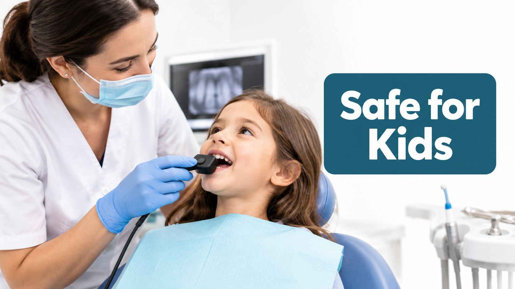
Dental X-rays are generally considered safe for children when clinically justified, with a typical dental X-ray delivering approximately 0.04 millisieverts (mSv), or 4 millirems, of radiation. The more useful question for a parent is which image this child needs, why it's needed now, and how the dental team will limit exposure.
A child may be sitting through a first cleaning in Amanda, Lancaster, Circleville, or Carroll when a hygienist mentions dental X-rays. The parent's mind can move quickly from “Is there a cavity?” to “Is this radiation safe?” That concern deserves a clear answer, not a reflexive yes or no.
Amanda Family Dental serves families seeking a dentist near me, a dentist in Amanda, OH, a dentist in Lancaster, OH, a dentist in Circleville, OH, or a dentist in Carroll, OH. For children, the central decision isn't whether every X-ray should be accepted or refused. It's whether the expected diagnostic benefit justifies a particular image for that child at that point in development.
Table of Contents
- When the Dental Chair Raises a Parent's Question
- How Much Radiation a Dental X-Ray Actually Delivers
- How ALARA Protects Children in the Dental Office
- Choosing the Right Type of Image for the Right Child
- How Often Kids Need X-Rays Based on Their Own Risk
- Why Skipping X-Rays Can Sometimes Be the Bigger Risk
- Smart Questions to Ask Before Your Child's Next Image
- What a Pediatric First Visit at Amanda Family Dental Looks Like
When the Dental Chair Raises a Parent's Question
A hygienist may mention an image while your child is in the chair for a cleaning. The parent's next question is often, “Can this wait?” It is a sensible question because children are more sensitive to ionizing radiation and have more years ahead in which potential effects could develop.
The American Academy of Pediatric Dentistry endorses ALARA, meaning exposure should remain “as low as reasonably achievable.” Its guidance recommends radiographs when the information could affect care, rather than using one schedule for every child (AAPD guidance on prescribing pediatric radiographs).
Before agreeing, ask what the image is meant to find: decay between the back teeth, an erupting permanent tooth, an injury, an infection, or an area the dentist cannot examine well. The answer should connect the image to a decision, such as whether treatment is needed or whether a developing tooth needs monitoring.
A child with visible tooth surfaces and a low risk of decay may need fewer images than a child with tight contacts, previous cavities, orthodontic concerns, pain, or recent trauma. Previous radiographs may also answer the question, so the dental team should review them before ordering another image.
The child's examination and dental history provide the context. Tooth development, symptoms, medical history, and the quality of earlier images all help determine which image fits this child, at this visit, and for this reason.
At Amanda Family Dental, parents can discuss the imaging plan during a new patient examination or preventive dental care visit. The team can explain what the image should show, how the equipment is used, and how unnecessary exposure is avoided. A calm explanation makes the decision easier to understand, without reducing it to a generic yes or no.
How Much Radiation a Dental X-Ray Actually Delivers
A parent may see several dose figures and wonder why they do not match. The U.S. Environmental Protection Agency lists a dental X-ray at approximately 0.04 mSv, or 4 millirems. The same source reports common intraoral dental radiographs at roughly 1.5 microsieverts and panoramic examinations at approximately 2.7 to 24 microsieverts (EPA information about radiation and medical imaging).
The units measure radiation dose at different scales. The 0.04 mSv figure reflects a full series of images, while the 1.5 microsievert figure reflects a single intraoral image, which is why the numbers differ. A full series includes more than one exposure, so it should not be compared directly with one bitewing or periapical image.
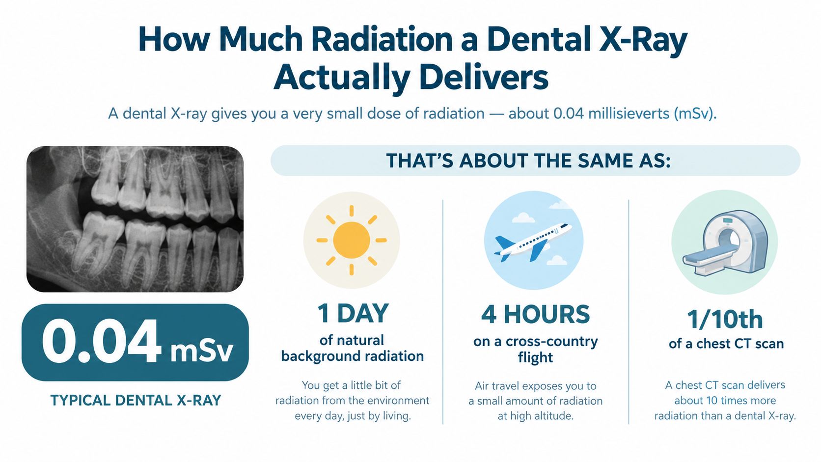
The useful question is not whether every image has the same dose. It is which image answers the child's dental question, and whether the result could change care. Routine dental images use a small amount of radiation, but children still benefit from avoiding images that do not have a clear purpose.
Routine images and advanced scans are different
A bitewing image shows the contact areas between back teeth, where decay may be hidden from a visual examination. A panoramic image gives a broad view of the teeth and jaws. These conventional images serve different purposes, and both generally involve less radiation than dental cone-beam computed tomography, commonly called CBCT.
CBCT produces a three-dimensional view. It may help when two-dimensional images cannot answer the diagnostic question, but three-dimensional technology alone is not a reason to use it. The U.S. Food and Drug Administration recommends dental CBCT only when conventional radiographs or other imaging cannot provide the information needed for diagnosis or treatment planning (FDA guidance on dental CBCT).
The FDA also recommends the smallest suitable field of view and exposure settings that preserve adequate image quality. For a child, more detail is useful only when it helps answer a specific question.
Dose is one part of the decision. The image type, area examined, child's risk, and likelihood of changing treatment all matter.
How ALARA Protects Children in the Dental Office
ALARA becomes practical in the dental chair. A family dentist in Amanda, OH, should be able to explain four decisions: which equipment the office uses, how exposure is set, how the child is positioned, and why this image is needed now. The goal is a useful image with no unnecessary exposure.
The receptor matters
A fast image receptor needs less exposure to produce a diagnostic image. Pediatric radiography guidance reports that moving from slower D-speed film to F-speed film or digital receptors can reduce dose by at least a factor of 2, according to the AAPD guidance cited earlier.
Digital sensors also show the image promptly. The dental team can check whether it answers the clinical question while the child is still in the chair. That immediate review may reduce repeats caused by processing problems or an image that was not captured clearly.
The beam should fit the image
Collimation narrows the X-ray beam to the area being examined. Rectangular collimation can reduce dose by approximately 4 to 5 times without harming image quality, according to the pediatric radiography guidance cited above.
The sensor size, the child's position, and the exposure setting must match the examination. A sensor that shifts, or a child who cannot remain still, may produce an image that does not answer the question. Repeating it adds exposure without adding useful information.
Protection and prescribing complete the process
The FDA lists safeguards that include using the fastest compatible receptor, matching the beam to the receptor, applying suitable exposure and processing techniques, and using protective aprons and thyroid collars when appropriate (FDA guidance on selecting patients for dental radiographic examinations).
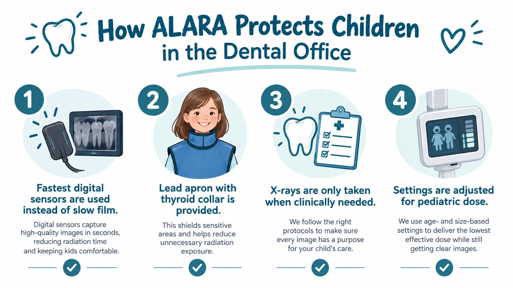
Parents can ask:
- Digital imaging: Does the office use a fast receptor suited to children?
- Child-sized settings: Will the exposure be adjusted for the child?
- Focused beam: Will collimation match the image receptor?
- Clinical reason: What finding should this image evaluate?
The team should also review available prior images before ordering another one. If an earlier image already answers the question, a repeat adds exposure without clinical value. These checks make the decision specific to the child, the image, the interval, and the reason for imaging.
Choosing the Right Type of Image for the Right Child
A child with a sore tooth after a fall needs a different image from a child being checked for hidden cavities. Dental X-rays are selected by the question the dentist needs to answer, the child's development, and whether a simpler view will provide enough information.
A bitewing image looks between the back teeth, where early decay may remain hidden during a visual examination. A periapical image focuses on one tooth and the surrounding area. It can help assess a root, possible infection, or an injury-related concern.
A panoramic image shows the mouth, jaws, and developing teeth in one broad view. It may help evaluate tooth eruption or other changes in the developing dentition. A panoramic image is not automatically needed at every routine visit.
CBCT creates a three-dimensional view and is generally used for specific, complex questions. Examples include locating an impacted tooth, assessing craniofacial structures, evaluating trauma, or planning treatment when two-dimensional images do not show the area clearly. As noted in the FDA recommendations for dental CBCT, the scan should answer a clinical question that simpler imaging cannot.
Pediatric dental imaging options at a glance
| Imaging type | Typical pediatric use | Approximate dose |
|---|---|---|
| Bitewing | Checking between back teeth for hidden decay | Common intraoral radiographs are reported at roughly 1.5 µSv |
| Periapical | Examining one tooth, its root, or a focused area | Dose varies with the examination and technique |
| Panoramic | Viewing the developing teeth and jaws broadly | Approximately 2.7 to 24 µSv |
| CBCT | Complex cases requiring three-dimensional information | Generally higher than conventional dental X-rays |
Parents can review digital X-ray advantages at Amanda Family Dental to learn more about current imaging technology. The practical question is simple: what will this image show that an examination or a simpler image cannot?
How Often Kids Need X-Rays Based on Their Own Risk
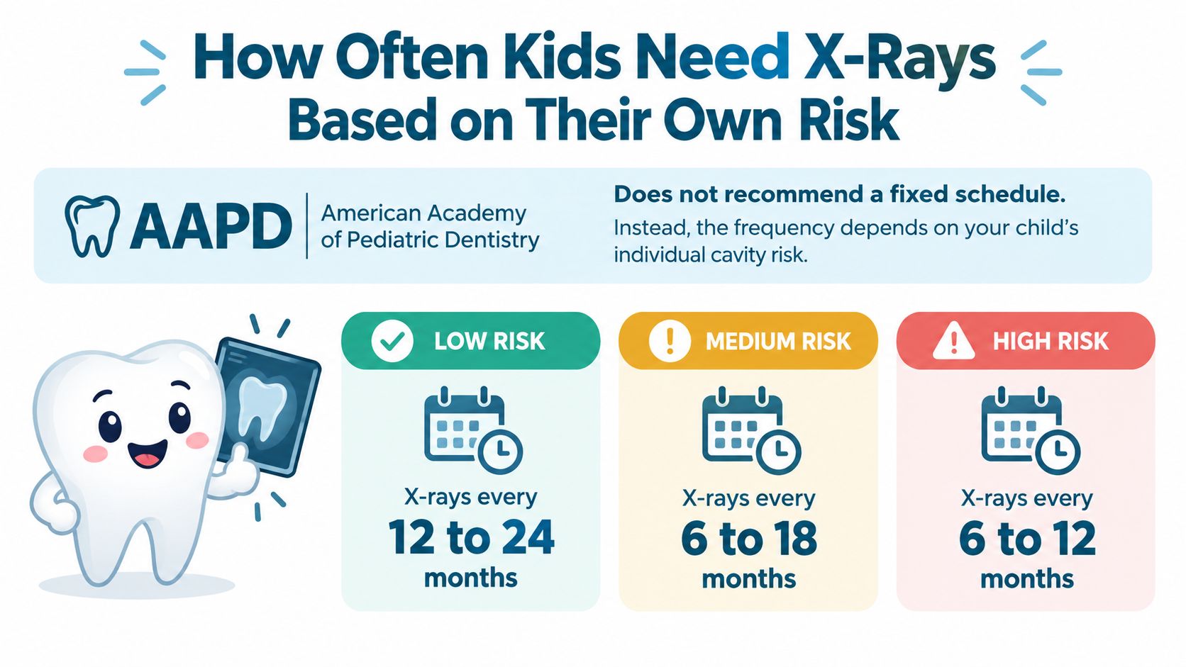
A calendar alone doesn't determine when a child needs dental X-rays. The American Academy of Pediatric Dentistry considers the child's age, tooth development, disease risk, examination conditions, and dental history. Its guidance gives different possible intervals rather than one schedule for every child (AAPD guidance on pediatric dental radiographs).
For example, posterior bitewings may be recommended at 6- to 12-month intervals for children with primary teeth when the contact surfaces can't be examined adequately. In some mixed-dentition situations, intervals may be 12 to 24 months, while other children may have intervals of 18 to 36 months or 24 to 36 months, depending on their circumstances.
Risk changes the interval
A child with a history of cavities, visible plaque, frequent exposure to sugary foods and drinks, or difficult-to-examine tooth contacts may need closer monitoring. A child with low decay risk and accessible tooth surfaces may need longer intervals.
The AAPD guidance notes that bitewing intervals may be roughly 2 to 3 years for children at low caries risk but about yearly for children at high risk. These ranges aren't promises or automatic prescriptions. They illustrate why a dentist should explain the reason behind a proposed interval.
A parent in Amanda, Lancaster, Circleville, or Carroll can ask:
- Risk assessment: What features of the child's examination or history affect the timing?
- Visibility: Can the relevant tooth surfaces be examined directly?
- Previous records: Could recent images from another dentist answer the question?
- Treatment impact: What decision would change if the image showed decay, delayed eruption, or another concern?
The guide to how often dental X-rays may be needed can help families prepare for that conversation. A child's imaging schedule should change when the child's risk changes, rather than repeating out of habit because a routine interval has passed.
Why Skipping X-Rays Can Sometimes Be the Bigger Risk
Avoiding unnecessary radiation is sensible. Refusing every clinically justified image isn't always the safer choice. Decay between tightly spaced teeth, infection inside a primary tooth, an unusual eruption pattern, or trauma affecting a developing permanent tooth may not be visible during a routine visual examination.
Consider a child whose back teeth touch tightly together. A dental probe may not reach the contact area, and the tooth may look normal from above. A bitewing image can reveal whether decay is present and help the dentist decide whether preventive care, a filling, or continued observation is appropriate.
A second example involves a front-tooth injury during play or sports. The visible crown may appear intact while the root or the developing permanent tooth requires evaluation. In that situation, a focused image can help the dentist assess the injury and plan care instead of waiting for pain, swelling, or a change in tooth color.
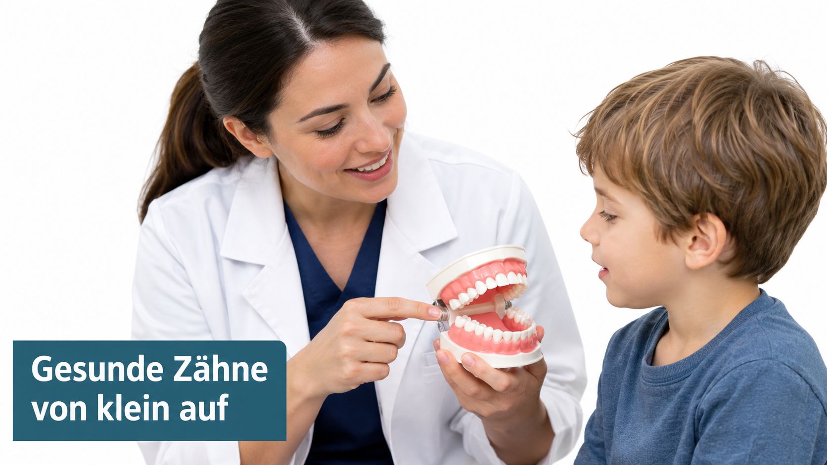
A well-timed image can prevent a child from needing more involved treatment later, but an image with no clear diagnostic purpose adds little value.
The balance is clinical reasoning. A dentist shouldn't order images automatically, and a parent shouldn't assume that avoiding every image eliminates risk. Finding hidden disease early can support pain relief, protect developing teeth, preserve chewing function, and reduce the chance that a small concern becomes an emergency dentist visit.
Smart Questions to Ask Before Your Child's Next Image
A parent can take part in the decision without knowing every imaging setting. The right questions connect the image to your child's specific situation: which image, for which child, at what interval, and why now?
- Ask what diagnosis the image could change: The answer should name a clinical question, such as hidden decay, eruption, trauma, infection, or an area the dentist cannot examine directly.
- Ask whether earlier images can answer that question: If useful records are available, the team should review them before taking a new image.
- Ask why the timing fits your child: The explanation may involve decay risk, development, symptoms, dental history, or today's examination.
- Ask whether the image is designed for children: The exposure settings and receptor should suit the child and the diagnostic purpose.
- Ask how the field is limited: Collimation keeps the beam focused on the area needed for the image.
- Ask what protective equipment is appropriate: A thyroid collar or lead apron may be considered when it does not interfere with the image.
The FDA dental radiography selection guidance supports prescribing dental radiographs according to the patient's clinical circumstances rather than following a fixed schedule.
At Amanda Family Dental, these questions can be part of the examination conversation. Ask what the image may show, how the result could affect care, and whether waiting would change the decision. A clear answer should help you understand why this image is being recommended for your child today, rather than treating imaging as an automatic part of every visit.
What a Pediatric First Visit at Amanda Family Dental Looks Like
A first visit should begin with a gentle visual examination and a conversation about the child's medical and dental history. The team can discuss tooth development, previous cavities, pain, trauma, brushing habits, and any concerns the parent or child has brought to the appointment.
If prior X-rays exist, the office can review them before considering new images. A digital image is prescribed only when the examination and history indicate that it could provide information needed for diagnosis or treatment planning. Digital sensors, child-sized exposure settings, and focused beam collimation support the ALARA approach described earlier.
The visit may also include preventive cleaning and exams, pediatric dental care, restorative treatment, or guidance about an urgent concern. Families who are preparing for a first appointment can review what to bring to a first dentist appointment, including relevant medical information and prior dental records.
Amanda Family Dental provides family and cosmetic dentistry for patients in Amanda, OH, Lancaster, OH, Circleville, OH, and Carroll, OH. A personalized plan can also address tooth extraction, restorative dentistry, dental implants near me, cosmetic dentistry, teeth whitening, or emergency dental services when those needs arise.
Schedule a child's examination with Amanda Family Dental to discuss dental X-rays, preventive care, and any concern about pain, eruption, decay, or injury. Contact the office to request an appointment or consultation in Amanda, Lancaster, Circleville, or Carroll, and bring any previous dental records so the team can make the most informed imaging decision.